Corneal Biopsy

What is a Cornea Biopsy?
Although far from a typical procedure, sometimes a corneal biopsy can be useful for more detailed information about the eye, especially when other treatments are not having their intended effect.
As with all tissue, a biopsy is simply the removal of a piece of tissue to use for testing. In this case, the tissue is taken from the cornea.
Who is a Candidate for a Corneal Biopsy?
A corneal biopsy is typically used as a diagnostic procedure in the management of complex keratitis. This is necessary if the patient has had a poor response to the usual antibiotics, if corneal scraping has failed to identify the organism, and if it has infiltrated deeper into the corneal stroma.
Keratitis is inflammation of the cornea, the clear, dome-shaped tissue on the front of the eye that covers the pupil and iris. Keratitis may be caused by an infection or an injury. Infectious keratitis can be caused by bacteria, viruses, fungi, and parasites. These are the cases that could merit a corneal biopsy to identify the cause of the infection.
Chronic keratitis caused by parasitic infections has become more of a problem beginning in the 1980s with the increasing popularity of soft contact lenses. Corneal biopsy has proven to be a dependable method of developing a definitive diagnosis in chronic cases of parasitic keratitis.
What Happens During a Corneal Biopsy?
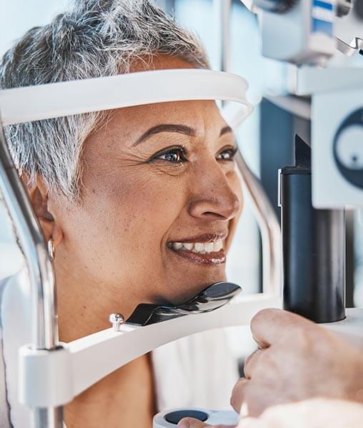
These are typically partial-thickness biopsies, meaning they do not penetrate through the full depth of the cornea. Partial-thickness samples should deliver the necessary information required to get at the root of the infection. They are performed while the patient is at the slit-lamp or operating microscope in our Center For Eye Care Excellence offices.
These are punch biopsies of up to 2 to 3 mm. The trephine is placed on the cornea and rotated between the thumb and forefinger to create a sample of usually only 1 mm. This piece is then grasped very carefully with toothed forceps so as not to crush the tissue. A crescent blade is then placed horizontally against the cornea to cut the tissue sample free. The biopsy specimen is transferred to the examining media. It can be cut into smaller pieces as necessary.
FAQ
Patient Testimonial
“Every time I have an appointment at Center For Eye Care Excellence, the entire staff makes me feel at ease and welcome. They take the time to listen to my concerns and worries. Even when the situation with my cornea was at it’s worst, Dr. Eden and all the staff made me feel comfortable and confident that they would do everything possible to help me. I never feel like “just another eye” but like they see me as a whole person who hopes to maintain her lifestyle and vision.”
– Kathleen
Schedule Your Consultation Today
If you’re interested in learning more about cornea biopsy please contact us for a consultation at 518.475.1515">518.475.1515 or fill out our contact us form below. We will discuss your needs and concerns, and determine your best course of action.

